


VIDEO: "Scientist Confirms White Clots Caused by Shots Are Infectious Amyloid" - Pub. May 8, 2025 by ForbiddenKnowledgeTV.net
Dr Kevin McCairn joins Maria Zeee with a high-level science presentation, with lots of electron microscope images of the mysterious white rubbery clots that morticians began discovering in the bodies of people all over the world after the COVID-19 injection mandates.
In Dr McCairn's analysis, these strange white clots are infectious amyloids and he hypothesizes that the SARS-CoV-2 spike protein present in both the 'viral agent' and the injections is a broad-acting amyloidogenic attack weapon.
He says the narratives about "viruses aren't real" and "the shots are full of nanobots" are being seeded by the perpetrators, who want to distract you from finding the data, "That they fired toxic amyloids at everyone. There's no little nanobots. I've shown that – I've scanned [with an electron microscope] dozens and dozens of vials. There's no graphene oxide. Nothing, OK? It's the toxic products of these programs that have done this. There is scientific data that you can follow, it is reproducible.
"We've got to find a solution to the amyloid problem and people have been trying to that for decades and we haven't made much progress."
He tells Maria Zeee:
"The important thing people need to take away from this interview is that these misfolded peptides, we now know are implicated, not just in neurodegeneration but also cancer.
"Because of this particular peptide being a blood peptide, it's going to be tied to cardiovascular and thrombotic events.
"And the disease burden – again, this is for somebody designing a weapon – it's dispersed, chaotic and most people are going to have a job tying it back to something as abstract as what I'm showing you on the screen, here."
Amyloids are formerly healthy proteins that have become degenerate and that are deposited as plaques in the brain, causing dementia.
Prions are infectious amyloids that induce surrounding proteins to likewise misfold and to degenerate, causing horrifying diseases, like Bovine Spongiform Encephalitis ("Mad Cow Disease") and its human analog, Creuzfeldt-Jakob.
All prions are amyloids but not all amyloids are prions. He says there's a spectrum of infectivity among prions, with scrapie being the most dangerous. There have been discussions as to whether the amyloids of Parkinsons and Azheimer's are infectious and whether there should be precautions taken during autopsies, etc. but the consensus, so far is just to "be careful".
Unlike other infectious agents, such as bacteria and fungi, prions do not contain DNA or RNA. Prions are proteins that infect other proteins, at the molecular level.
Dr McCairn says, "We're now going to be dealing with the consequences of the mass exposure to weaponized amyloids, either through the 'viral agent' or through...the 'vaccine' and the expression of the spike protein."
The next question is, would exposure to COVID clots be capable of seeding and catalyzing an amylodogenic cascade? and he says the preliminary data says "There seems to be something we should be looking at, right now."
Maria Zee and Dr McCairn are joined by Richard Hirschman, who was the first mortician to come forward with samples of these huge, rubbery COVID clots, such as these:
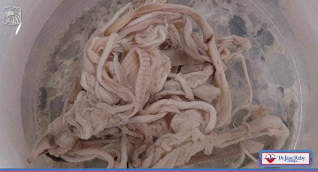
IMAGE 1: These white rubbery clots have been removed from from the bodies of those who've died from the COVID "Clotshot" injection, worldwide.
Dr McCairn cites the December 2021 scientific paper, "Subcutaneous Uptake on [18F]Florbetaben PET/CT: a Case Report of Possible Amyloid-Beta Immune-Reactivity After COVID-19 Vaccination" by Riccardo Laudicella et al., which he says should have set off alarm bells and stopped the vaccine roll-out, right then and there but he says many tenured and well-paid professionals knew better said nothing.
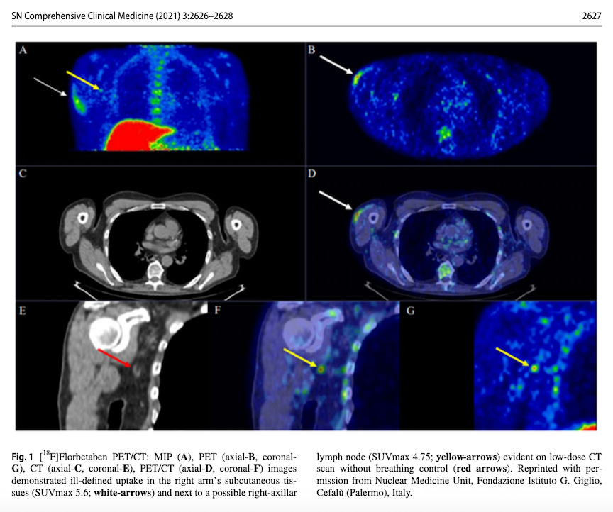
IMAGE 2: "Subcutaneous Uptake on [18F]Florbetaben PET/CT: a Case Report of Possible Amyloid-Beta Immune-Reactivity After COVID-19 Vaccination"
The in vivo study above involved the PET scan of a recently-vaccinated 70-year-old man, who was being tested for Alzheimer's disease. As part of the scan, he was administered Florbetaben, a diagnostic radiotracer, which lights up when it binds with β-amyloid plaques. This man's COVID injection site lit up, indicating that there was β-amyloid in the vaccine.
Dr McCairn did a study of Richard Hirshman's COVID injection clots to explore his hypothesis that our environment is being seeded with weaponized amyloids.
He took several images of a COVID clot at various degrees of magnification.
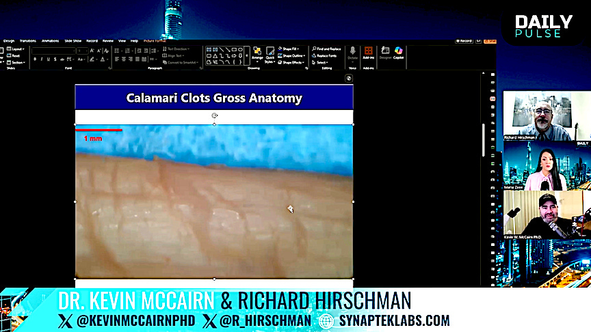
IMAGE 3: COVID amyloid clot at 1mm from Dr Kevin McCairn's presentation
He sliced the COVID clot tissue with a microtome, in order to be able to put extremely thin sections on slides for fluorescent microscopy.
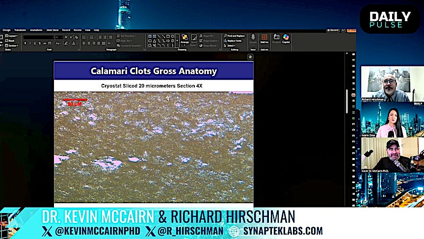
IMAGE 4: COVID amyloid clot sliced at 20 micrometers at 4X magnification from Dr Kevin McCairn's presentation
He was surprised to find that the COVID clot tissue was uniformly auto-fluorescent, which is not typical of normal human tissue, saying, "This, in and of itself was striking, this should have most scientists and pathologists who work in the domain thinking there was something important here to look at.
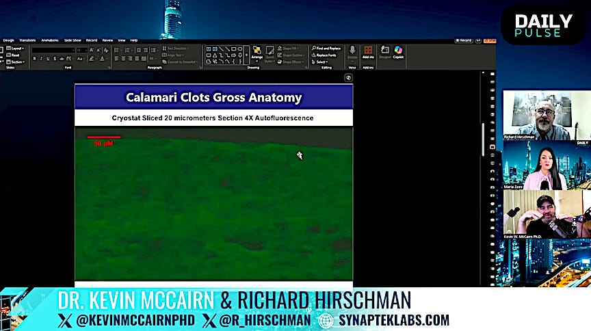
IMAGE 5: COVID amyloid clot sliced at 20 micrometers at 4X magnification from Dr Kevin McCairn's presentation showing autofluorescence.
"The amyloid microclots, which have been associated with viral infection, Long COVID and also vaxx injury, they're known to auto-fluoresce, as well but they're tiny, with respect to what you see here...we have this uniform, weird feature...If you found something like that under any other type of circumstance, this would be a whole avenue of research that a lab could take on. Why is there so much auto-fluorescence across this tissue?"
At 40X magnification, very dense fibrin protein structures were visualized, unlike anything he's ever seen before.
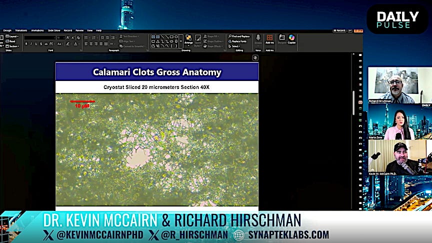
IMAGE 6: COVID amyloid clot sliced at 20 micrometers at 40X magnification from Dr Kevin McCairn's presentation showing very dense fibrin, unlike anything natural
Dye was then applied to the sample and it was found to be taken-up by certain regions in the COVID clot tissue, indicating the presence of an amyloid. Dr McCairn says this is something that people in his profession would normally want to investigate, especially as the detected amyloid/s could be infectious, like such as those that drive Creutzfeld-Jakob and Mad Cow Disease.
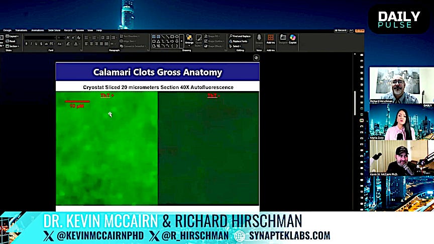
IMAGE 7: COVID amyloid clot sliced at 20 micrometers at 40X magnification, with and without dye from Dr Kevin McCairn's presentation
So, the COVID clot tissue is confirmed to contain amyloids. The next question is, are these amyloids associated with proteinopathy and neurogegenerative disease?
Below is an image of the COVID clot tissue, sliced even finer, taken with an electron miscroscope at 25X SEM:
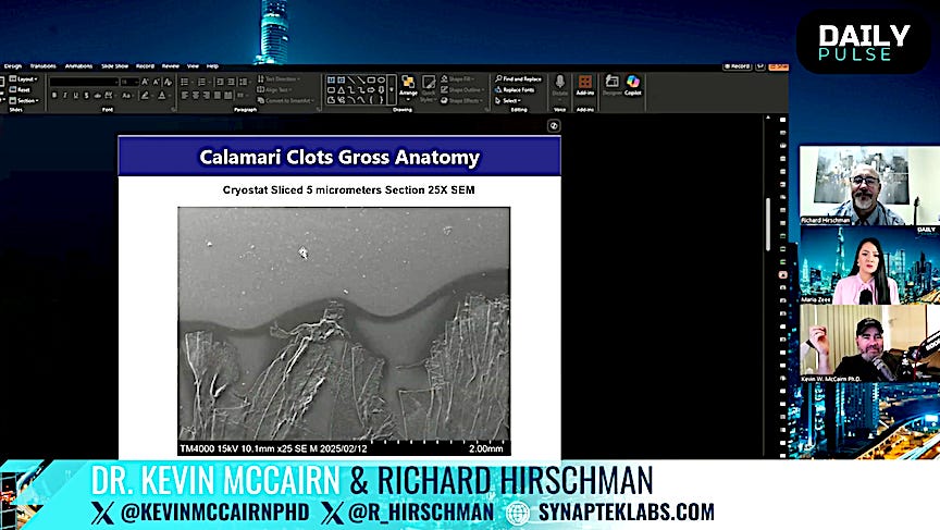
IMAGE 8: COVID amyloid clot sliced at 5 micrometers at 25X Scanning Electron Microscopy from Dr Kevin McCairn's presentation
Here's just one of the fibers above, visualized at 250X SEM, where you can see various features, like the "undulating topography":
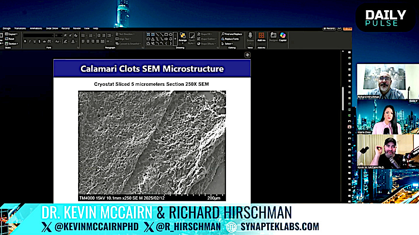
IMAGE 9: COVID amyloid clot sliced at 5 micrometers at 250X Scanning Electron Microscopy from Dr Kevin McCairn's presentation
Here's a visualization of the same fiber at 1000X SEM, revealing what are technically called "branching fibrils", the linear structures coming out of the globular forms and connecting to other globular structures.
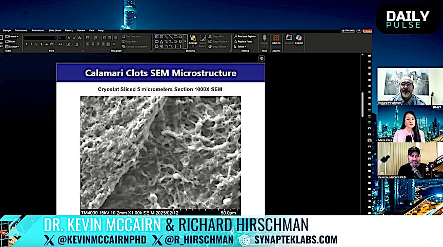
IMAGE 10: COVID amyloid clot sliced at 5 micrometers at 1000X Scanning Electron Microscopy from Dr Kevin McCairn's presentation
Below, is a visualization of the same fiber at 5000X SEM, with a closer view of the branching fibrils. This is the scale at which Dr McCairn explains that those in his field would be able to tell you what's unusual about this picture. He says, "Fibrin is a very long, 'noodle', spaghetti-like protein...It's very long, smooth-looking peptide and what you're looking at here doesn't look like that...[see Image 12 to compare this with the appearance of normal fibrin].
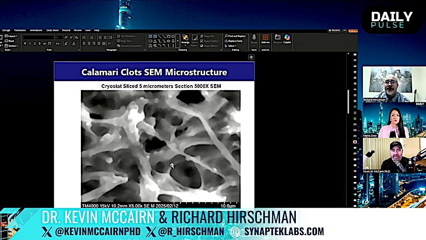
IMAGE 11: COVID amyloid clot sliced at 5 micrometers at 5000X Scanning Electron Microscopy from Dr Kevin McCairn's presentation
"Again, we can see these nodules that are probably what we would call 'nucleation points', where, perhaps branches are beginning to form and one of the stand-out features, immediately for me, which is why I like this image, as a scientist, if you look at...that branch and if you can see that it kind of has a rotation to it and it's twisted, what that tells us is the way that the protein is sort-of assembling itself has gone radically wrong.
"And in a sense, what's happening is, as the protein tries to build itself from its monomer units, they should come together and build the normal structures but in this instance, there's a torque being applied to the...amino acids of the monomer, coming together and as they join, they're being torqued and twisted and what that does is it changes the ability of other enzymes to come in, that would normally break the fibrin down...plasminogen is the primary one, in this instance.
"So the shape dictates the function in biology and the shape, here is radically wrong...You're looking at a very, very pathological, sick form of protein."
Dr McCairn cites the paper published last year, "Fibrin drives thromboinflammation and neuropathology in COVID-19", by Jae Kyu Ryu et al., which he encourages everyone to read, although it is highly-technical.
He says that what this paper is saying is, "That twisted peptide that I just showed you is attacking the brain, kind of in line with the hypothesis that you're dealing with these amyloidogenic attack weapons.
"And the way to do that, in this instance is a broad-acting weapon that's acting as a protein that's in very, very large amounts in the blood that is...able to cause inflammation in the brain and presumably...be seeding more amyloid into the brain.
The image below repeats the image above with inset electron microscope images from the above paper of human blood plasma. The image on the left shows normal fibrin, which looks smooth, next to an image of blood plasma that's been exposed to the spike protein that is a feature of both the SARS-CoV-2 virus and of the COVID-19 injections, in which the fibrin, which has similar nodules to those seen in the electron microscope image if the COVID clot tissue.
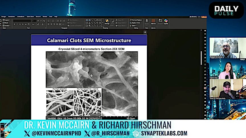
IMAGE 12: COVID amyloid clot sliced at 5 micrometers at 5000X Scanning Electron Microscopy with images of normal human blood plasma and plasma exposed to the SARS-CoV-2 spike protein from Dr Kevin McCairn's presentation
The image of exposed blood plasma from the above paper, in the inset image doesn't look as bad as the sample of COVID clot tissue obtained by Richard Hirschman but there are similarities.
Dr McCairn explains that, because the body doesn't see amyloids as foreign, it doesn't produce antibodies for them and that's why they accumulate and penetrate organ systems and cause disease.
As for the infectivity of the COVID amyloid, there's an assay technique called Real-Time Quaking-Induced Conversion, aka RT-QuIC, which is used to detect Creuzfeldt-Jakob disease.
Dr McCairn used this technique, taking a seed-sized portion of the COVID clot provided by Richard Hirschman and he "challenged" it with human plasma, shaking it for 7.5 hours. There was a strong reaction, which produced an increasing amount of misfolded amyloid cores, lighting-up with increased fluorescence, similar to that seen in Images 5 and 7, above.
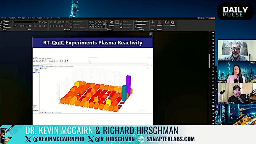
IMAGE 13: Result of RT-QuIC assay of COVID amyloid clot from Dr Kevin McCairn's presentation
The next question is, "Is it capable of seeding and catalyzing an amyloidogenic cascade?" and he says the preliminary data says there seems to be something we should be looking at, right now.
McCairn says that, "The fact that this is being ignored by authorities is very disturbing, because...so many people have been exposed...that I would say that all the blood supply is probably contaminated, with respect to hospitals, etc.
"And what would it mean, with respect to [potential COVID amyloid] transmission between person-to-person?...It's likely to come down to tissue exposure. I don't think just bumping into someone is going to cause the transmission chain to continue."
Maria confirms that we're not certain whether COVID clots are prions and infectious but she asks him what would be the implications if they are, noting that one of his slides (not shown here) is entitled "Super Prion Event".
He replies that a Super Prion Event has been put forward as the cause of the Neanderthal extinction, whereby humans spread a kuru-type of disease to Neanderthals, who were less immune, which also suggests that cannibalism was once widespread among humans.
He's certain that the COVID clots are amyloidogenic, saying there is pushback to this in the medical community but he suspects there will now likely be less so, with USAID being defunded and dismantled.
He asks, "Is it as infective as the prion protein? I don't know. It's tough to say. I don't think so, to tell the truth but I don't think it's benign, either...
"These are known as surreptitious disease mechanisms, which means they take a long time to express themselves. When they do express themselves, acting at this fundamental substrate layer, it's very difficult to treat."
He says until now, this kind of illness was relegated to the elderly but there are now signals of this happening in clusters among the young and that our representatives need to be held culpable, at this point, although he doubts they will be.
To go deeper into Dr McCairn's research, visit his site, Synapteklabs.com, where he also offers to investigate samples to determine the amyloid burden of the donor from his laboratory in Gifu, Japan. His Twitter account is @KevinMcCairnPhD.